Moving From Film X-Ray to PACS: A Hospital Implementation Guide
Printed film still runs the radiology department in many hospitals in Pakistan: films are printed for every study, carried between departments, stored in envelopes and occasionally lost. A PACS (Picture Archiving and Communication System) replaces that with a digital archive and a viewer that authorised doctors can open from any workstation. This guide sets out the steps of a typical move from film to PACS, and what to check at each one.
Why Hospitals Move Off Film
The case for PACS usually rests on four things:
- Cost. Film, chemicals, envelopes and CDs are a recurring expense on every study. Reducing printing is often the largest single saving.
- Speed. A study is available to the radiologist as soon as the scan is complete, rather than after printing and delivery.
- Access. Referring doctors and other sites can view images and reports without anyone carrying films or CDs.
- Safety. A backed-up digital archive removes the risk of lost or damaged films, and prior studies are always available for comparison.
To put a figure on the first point for your own facility, the PACS ROI calculator estimates your current film spend and the savings from printing less.
1. Assess Your Imaging Estate
Start with an inventory. For every imaging device, record:
- the modality type, manufacturer and model;
- whether it outputs DICOM, and which DICOM options are enabled;
- monthly study volume, and how many studies are printed today;
- its location, and the network connection available at that location.
The inventory decides how much work each room needs before it can send images to PACS:
| What you have | What it needs to connect to PACS |
|---|---|
| CT, MRI, digital radiography (DR), digital mammography | Usually DICOM-ready: configure PACS as a DICOM storage destination. |
| Analog (film-screen) X-ray | A digitiser first: a computed radiography (CR) reader that scans reusable imaging plates, or a DR detector retrofit. |
| Ultrasound | Most current units support DICOM, but the DICOM option may need to be licensed or enabled by the vendor. |
| Older devices without DICOM | Check with the vendor for a DICOM upgrade; otherwise a capture or conversion device may be needed. |
The monthly volumes also give you a storage estimate: multiply studies per month by a typical study size for each modality, then by the number of years you need to keep online.
2. Choose On-Premise, Cloud or Hybrid
An on-premise PACS keeps servers and storage in your hospital, which suits facilities that must keep all data in-house and have IT staff to run them. A cloud PACS avoids a large upfront hardware purchase and grows with study volume, but depends on a reliable internet connection. A hybrid setup keeps a local cache in the hospital for fast viewing, with the archive in the cloud. Pacslink offers all three; the right choice depends on your internet reliability, IT staffing and budget.
3. Prepare the Network and Reading Stations
- Network. Each modality needs a wired network connection to the PACS (or to the local cache in a hybrid setup). Large CT and MRI studies are slow on weak links, so check the path from the busiest scanners first.
- Internet. For cloud or hybrid PACS and remote reading, plan for the upload capacity your study volume needs, and consider a backup connection.
- Power. Put servers, network switches and reading workstations on UPS so that outages do not interrupt image transfer or reporting.
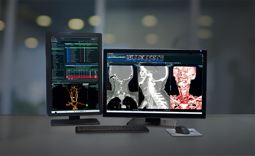
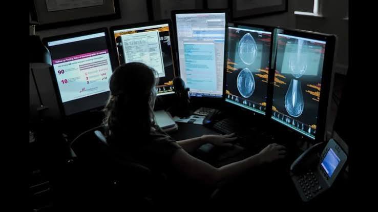
- Reading stations. Radiologists reporting primary diagnoses need suitable workstations and displays. Clinicians reviewing images and reports can use standard PCs, because the viewer runs in the browser.
4. Connect the Modalities
Each modality is configured once, usually with your biomedical engineer or the modality vendor's engineer:
- Give the modality an AE title (its DICOM name) and record its IP address and port.
- Add the PACS as a storage destination on the modality.
- If you have a RIS, add it as the modality worklist provider, so the scheduled patient appears at the console and nobody re-types demographics.
- Run a Verification (C-ECHO) test in both directions, then send a test study and check it in the viewer.
Our DICOM and HL7 integration standards guide explains these DICOM services in more detail.
5. Integrate With RIS and HMIS
PACS stores images; a Radiology Information System (RIS) manages the orders, schedules, worklists and reports around them, and the HMIS holds the patient record. Connected over HL7, a patient registered once in the HMIS flows through radiology without re-typing, and the signed report returns to the patient record. The HMIS and PACS integration guide walks through that workflow step by step.
6. Decide What to Do With Historical Images
- Digital studies already stored on modality hard drives or CDs can usually be imported into PACS as DICOM, so priors appear next to new studies.
- Printed films stay in the physical archive for their retention period. Scanning old films is rarely worth it except for specific cases.
- Agree a cut-off date and which priors matter clinically, for example recent studies of patients under follow-up.
7. Train Staff and Run a Pilot
Train each group on what changes for them: technologists on worklists and sending studies, radiologists on the viewer and reporting, and referring doctors and wards on finding images and reports. Start with one or two modalities, check that every study arrives complete and on the right patient, then add the rest. Keep printing in parallel during the pilot, and reduce it as doctors become comfortable viewing on screen.
8. Go Live and Measure
Track a few numbers from the first week, and compare them with your pre-PACS baseline:
- films printed per month, and film spend;
- time from scan to signed report;
- studies that arrive with missing images or on the wrong patient, which should fall to zero once worklists are in use;
- requests for re-scans because films were lost.
Readiness Checklist
- Modality inventory complete, with DICOM capability confirmed for each device
- CR or DR digitisers planned for every analog X-ray room
- Hosting model chosen and storage estimated
- Network, internet and UPS checked at every modality and reading location
- RIS / HMIS integration scope agreed
- Historical-image cut-off agreed
- Pilot modality, training plan and parallel-print period agreed
- Baseline figures recorded: film spend, report turnaround, re-scans
Every hospital's starting point is different. A site assessment turns this checklist into a timeline and cost for your facility; ask our Lahore team to arrange one.